
Page 197 - Edited - Webster HEAD AND NECK - part 1
P. 197
HN 163
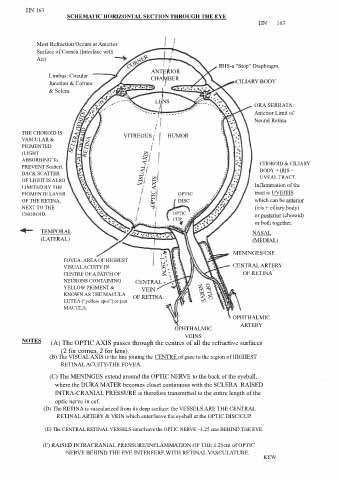
SCHEMATIC HORIZONTAL SECTION THROUGH THE EYE
HN 163
Most Refraction Occurs at Anterior
Surface of Cornea (Interface with
Air)
IRIS-a “Stop” Diaphragm.
ANTERIOR
Limbus: Circular CHAMBER
Junction & Cornea CILIARY BODY
& Sclera.
LENS
ORA SERRATA:
Anterior Limit of
Neural Retina.
THE CHOROID IS VITREOUS HUMOR
VASCULAR &
PIGMENTED
(LIGHT
ABSORBING To CHOROID & CILIARY
PREVENT Scatter). BODY + IRIS =
BACK SCATTER UVEAL TRACT.
OF LIGHT IS ALSO
LIMITED BY THE Inflammation of the
PIGMENTE LAYER OPTIC tract is UVEITIS
OF THE RETINA, DISC which can be anterior
NEXT TO THE (iris + ciliary body)
CHOROID. OPTIC or posterior (choroid)
CUP
or both together.
TEMPORAL NASAL
(LATERAL) (MEDIAL)
MENINGES/CSF.
FOVEA: AREA OF HIGHEST
VISUAL ACUITY IN CENTRAL ARTERY
CENTRE OF A PATCH OF OF RETINA
NEURONS CONTAINING CENTRAL
YELLOW PIGMENT & VEIN
KNOWN AS THE MACULA OF RETINA.
LUTEA (“yellow spot”) or just
MACULA.
OPHTHALMIC
ARTERY
OPHTHALMIC
VEINS
NOTES (A) The OPTIC AXIS passes through the centres of all the refractive surfaces
(2 for cornea, 2 for lens).
(B) The VISUAL AXIS is the line joining the CENTRE of gaze to the region of HIGHEST
RETINAL ACUITY-THE FOVEA.
(C) The MENINGES extend around the OPTIC NERVE to the back of the eyeball,
where the DURA MATER becomes closet continuous with the SCLERA. RAISED
INTRA-CRANIAL PRESSURE is therefore transmitted to the entire length of the
optic nerve in csf.
(D) The RETINA is vascularized from its deep surface: the VESSELS ARE THE CENTRAL
RETINAL ARTERY & VEIN which enter/leave the eyeball at the OPTIC DISC/CUP.
(E) The CENTRAL RETINAL VESSELS enter/leave the OPTIC NERVE ~1.25 cms BEHIND THE EYE.
(F) RAISED INTRACRANIAL PRESSURE/INFLAMMATION OF THE 1.25cm of OPTIC
NERVE BEHIND THE EYE INTERFERE WITH RETINAL VASCULATURE.
KEW
.