
Page 96 - Edited - Webster HEAD AND NECK - part 2-Merge PDF
P. 96
HN 289
LARYNGOSCOPY
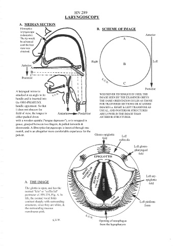
A. MEDIAN SECTION
Fibreoptics B. SCHEME OF IMAGE
laryngoscope
(schematic). Anterior
The tip would A
be advanced
until the best
view was
obtained.
Right Left
Anterior B
A
B
C
Posterior
C
Posterior
A laryngeal mirror is A
attached at an angle to its WHICHEVER TECHNIQUE IS USED, THE
handle and is inserted into B IMAGE SEEN BY THE EXAMINER OBEYS
the ORO-PHARYNX C THE SAME ORIENTATION RULES AS THOSE
FOR TRANVERSE SECTIONS OR SCANNED
handle uppermost. So that IMAGES i.e. RIGHT & LEFT TRANSPOSE AS
it does not obscure the USUAL, AND POSTERIOR STRUCTURES
field of view, the tongue is Anterior Posterior ARE LOWER IN THE IMAGE THAN
either pushed down ANTERIOR STRUCTURES.
with a wooden spatula ("tongue depressor"), or is wrapped in
gauze, grasped between two fingers, & pulled forwards &
downwards. A fibreoptics laryngoscope is inserted through one
nostril, and is an altogether more comfortable experience for the
patient. Glosso-epiglottic Left
fold vallecula
Left glosso-
pharyngeal
fold
EPIGLOTTIS
Left ary-
A. THE IMAGE epiglottic
fold
GLOTTIS
The glottis is open, and has the
normal "kite" or "coffin lid"
perimeter cf. HN 274, Fig. A. In
life, the normal vocal folds
contrast sharply with surrounding Left piriform
structures, since they are white, & fossa
the surrounding mucous
membranes pink.
Opening of oesophagus
from the hypopharynx