Page 64 - Edited - Webster HEAD AND NECK - part 2-Merge PDF
P. 64

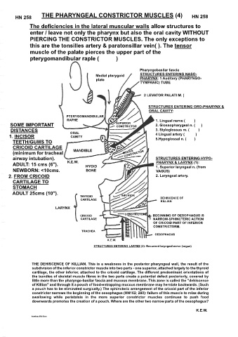
THE PHARYNGEAL CONSTRICTOR MUSCLES (4)
         ΗΝ 258                                                                                  ΗΝ 258
                The deficiencies in the lateral muscular walls allow structures to
                enter / leave not only the pharynx but also the oral cavity WITHOUT
                PIERCING THE CONSTRICTOR MUSCLES. The only exceptions to
                this are the tonsilles artery & paratonsillar vein( ). The tensor
                muscle of the palate pierces the upper part of the
                pterygomandibular raple (           )


                                                                       Pharyngobasilar fascia
                                                 Medial pterygoid      STRUCTURES ENTERING NASO-
                                                 plate                 PHARYNX: 1 Auditory (PHARYNGO-
                                                                       TYMPANIC) TUBE


                                                                         2 LEVATOR PALATI M. (


                                                                           STRUCTURES ENTERING ORO-PHARYNX &
                                                                           ORAL CAVITY:
                                  PTERYGOMANDIBULAR
                                  RAPHE                                        1. Lingual nerve (         )
      SOME IMPORTANT                                       SUPERIOR            2. Glossopharygeal n. (      )
                                                           CONSTRICTOR
      DISTANCES                     ORAL                                       3. Styloglossus m. (         )
      1. INCISOR                    CANITY          8                          4 Lingual artery (           )
                                                                               5.Hypoglossal n. (        )
       TEETH/GUMS TO
       CRICOID CARTILAGE
       (minimum for tracheal          MANDIBLE
       airway intubation).                                                     STRUCTURES ENTERING HYPO-
       ADULT: 15 cms (6").         K.E.W.   HYOID                              PHARYNX & LARYNX (1):
                                                                               1. Superior laryngeal n. (from
       NEWBORN: <10cms.                     BONE                               VAGUS)
     2. FROM CRICOID                                                           2. Laryngeal artery.
       CARTILAGE TO
       STOMACH
       ADULT 25cms (10").
                                         THYROID              INFERIOR           DEHISCENCE OF
                                         CARTILAGE
                                                                                 KILLIAN
                             LARYNX
                                                               CONSTRICTOR
                                         CRICOID                              BEGINNING OF OESOPHAGUS IS
                                         CARTILAGE                            NARROW-SPHINCTERIC ACTION
                                                                              OF CRICOID PART OF INFERIOR
                                                                              CONSTRICTORM.
                                          TRACHEA
                                                                              OESOPHAGUS
                                                       K.E.W.
                                                STRUCTURES ENTERING LARYNX (2): Recurrent laryngeal nerve (vagus)


                 THE DEHISCENCE OF KILLIAN. This is a weakness in the posterior pharyngeal wall, the result of the
                 subdivision of the inferior constrictor muscle into two parts - one superior, attached largely to the thyroid
                 cartilage, the other inferior, attached to the cricoid cartilage. The different predominant orientations of
                 the bundles of skeletal muscle fibres in the two parts create a potential defect posteriorly, covered by
                 little more than the pharyngo-basilar fascia and mucous membrane. This zone is called the "dehiscence
                 of Killian" and through it a pouch of food-entrapping mucous membrane may herniate backwards. (Such
                 a pouch has to be eliminated surgically.) The sphincteric arrangement of the cricoid part of the inferior
                 constrictor narrows the beginning of the oesophagus (HN162; 280): failure of this muscle to relax during
                 swallowing  while  peristalsis  in  the  more  superior  constrictor  muscles  continues  to  push  food
                 downwards promotes the creation of a pouch. Where are the other two narrow parts of the oesophagus?

                                                                                                   K.E.W.
                 kewteach\killian
   59   60   61   62   63   64   65   66   67   68   69